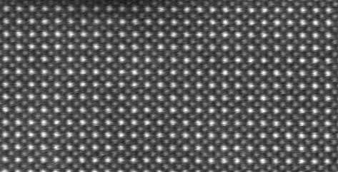
Crystal structure
Ordered arrangement of particles in crystalline materials.

Biochemlife · CC BY-SA 4.0
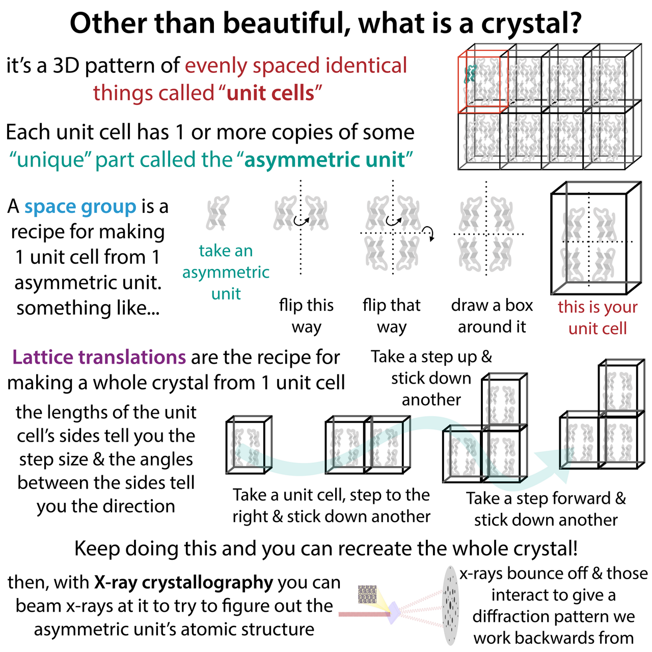
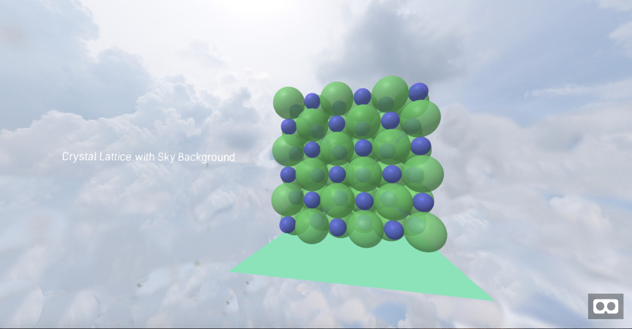
Crystal structure is a description of the ordered arrangement of atoms, ions, or molecules in a crystalline material. Ordered structures occur from the intrinsic nature of constituent particles to form symmetric patterns that repeat along the principal directions of three-dimensional space in matter. The smallest group of particles in a material that constitutes this repeating pattern is the unit cell of the structure, which completely reflects the symmetry and structure of the entire crystal.
- field
- Crystallography
- known_for
- Description of ordered atomic arrangements, unit cells, Bravais lattices, space groups, Miller indices, and interplanar spacing formulas
Lore & Background
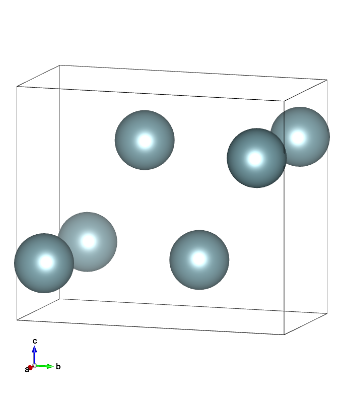
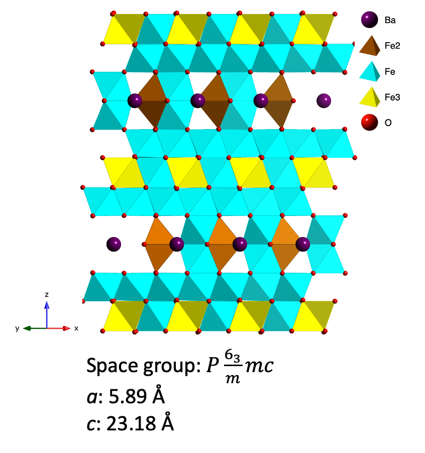
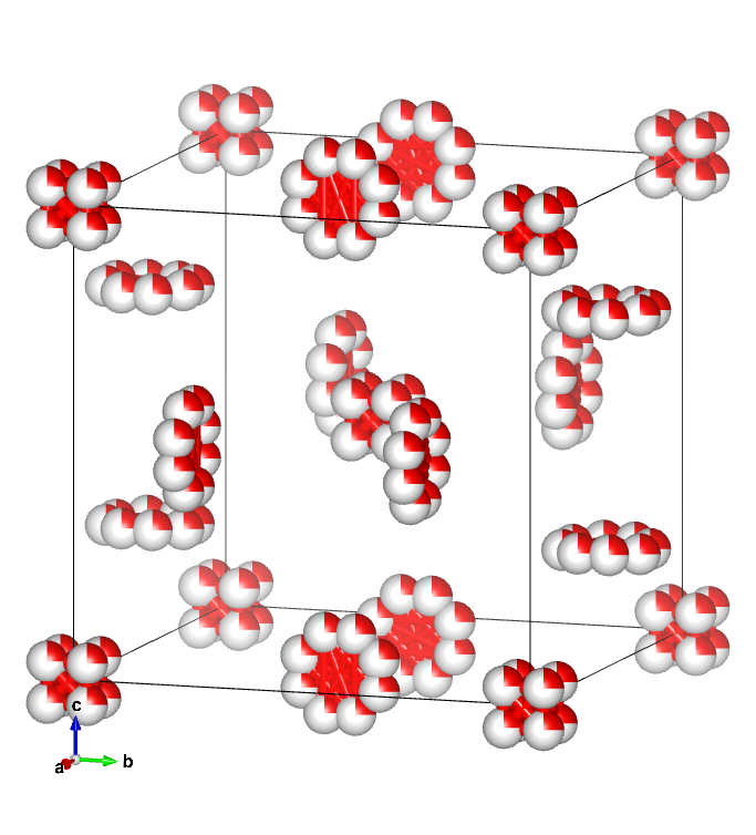
Crystal structure is described in terms of the geometry of the arrangement of particles in the unit cells. The unit cell is defined as the smallest repeating unit having the full symmetry of the crystal structure. The geometry of the unit cell is defined as a parallelepiped, providing six lattice parameters taken as the lengths of the cell edges (a, b, c) and the angles between them (α, β, γ). The positions of particles inside the unit cell are described by the fractional coordinates (xi, yi, zi) along the cell edges, measured from a reference point. The asymmetric unit may be chosen so that it occupies the smallest physical space, and all other particles of the unit cell are generated by the symmetry operations that characterize the symmetry of the unit cell. The collection of symmetry operations of the unit cell is expressed formally as the space group of the crystal structure.
Reader's Guide
Crystal structure and symmetry play a critical role in determining many physical properties, such as cleavage, electronic band structure, and optical transparency. Vectors and planes in a crystal lattice are described by the three-value Miller index notation. The syntax (hkℓ) denotes a plane that intercepts the three points a1/h, a2/k, and a3/ℓ, or some multiple thereof. The Miller indices for a plane are integers with no common factors. Some directions and planes have a higher density of nodes, influencing optical properties, adsorption and reactivity, surface tension, microstructural defects, cleavage, and plastic deformation. For cubic crystals with lattice constant a, the spacing d between adjacent (ℓmn) lattice planes is given by d = a / √(ℓ² + m² + n²). Coordinates in angle brackets such as ⟨100⟩ denote a family of directions that are equivalent due to symmetry operations, and coordinates in curly brackets such as {100} denote a family of plane normals that are equivalent due to symmetry operations.
Did You Know?
- The smallest repeating unit having full symmetry of the crystal structure is the unit cell, defined as a parallelepiped with six lattice parameters.
- Miller indices (hkℓ) denote a plane that intercepts the three points a1/h, a2/k, and a3/ℓ, or some multiple thereof.
- For cubic crystals, the spacing d between adjacent (ℓmn) lattice planes is given by d = a / √(ℓ² + m² + n²).
The Core Principle: Turning Invisible Atoms into Measurable Geometry
The fundamental idea behind X-ray crystallography is that a crystal's internal atomic arrangement acts as a natural diffraction grating for incoming X-rays. When the beam strikes the ordered lattice, radiation scatters in specific directions dictated by the spacing and orientation of the atoms. A crystallographer records the angles and intensities of those scattered beams and then reconstructs a three-dimensional map of electron density. From that density map, the positions of individual atoms, the character of their chemical bonds, and any crystallographic disorder present in the sample can all be extracted. This makes the technique uniquely powerful: it converts an invisible atomic architecture into measurable geometric data, yielding a level of structural detail that no other single experimental method can provide so completely. The result is a comprehensive picture of matter at the scale where chemistry and physics meet.
A Long Prehistory of Crystallographic Thought
Long before X-rays entered the picture, scientists had been puzzling over why crystals display such striking geometric regularity. In 1611, Johannes Kepler mused that the hexagonal shape of snowflakes arose from a regular packing of tiny spherical water particles. Decades later, Nicolas Steno demonstrated experimentally that the angles between faces remain constant for a given crystal type, a principle that still underpins the field. René Just Haüy pushed further in 1784, showing that every crystal face could be described as a simple stacking of identical blocks, a law of decrements that implicitly pointed toward an underlying three-dimensional periodic array. William Hallowes Miller then formalized face identification in 1839 with his system of three small integers, the Miller indices, which remain standard today. The full catalog of possible crystal symmetries was assembled in the nineteenth century by Hessel, Bravais, Fedorov, Schönflies, and, somewhat belatedly, William Barlow, whose 1880s structural proposals would later be confirmed by X-ray evidence.
From Röntgen's Mystery to von Laue's Spots
Wilhelm Röntgen's 1895 discovery of X-rays left the physics community uncertain about what they actually were. William Henry Bragg argued in 1907 that they could not be electromagnetic radiation, pointing to their ability to ionize gases. The wave interpretation gained support when Charles Glover Barkla demonstrated transverse polarization and sharp spectral lines, and when single-slit work in Arnold Sommerfeld's lab estimated a wavelength near one angstrom. The decisive moment arrived in 1912, when Paul Peter Ewald and Max von Laue, chatting in Munich's English Garden, realized that a crystal's unit-cell spacing might match the X-ray wavelength well enough to produce diffraction. Von Laue, with technicians Walter Friedrich and Paul Knipping, passed a beam through a copper sulfate crystal and captured a photographic plate showing a striking pattern of spots arranged in intersecting circles. Presented to the Bavarian Academy that June, the result confirmed X-rays as electromagnetic waves and earned von Laue the 1914 Nobel Prize in Physics.
From the Laboratory to the Clinic
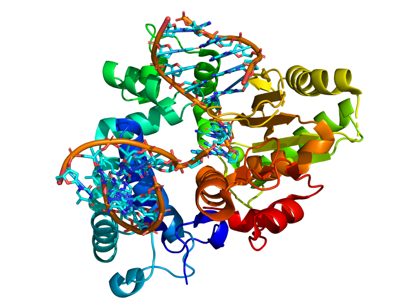
X-ray crystallography has shaped nearly every branch of science that deals with matter at the atomic scale. In its earliest decades it pinned down atomic sizes, bond lengths, and the subtle structural differences between minerals and alloys. In biology it has revealed the three-dimensional shapes of vitamins, therapeutic drugs, proteins, nucleic acids including DNA, and even whole viruses, linking molecular architecture to biological function. Today it remains the principal tool for distinguishing materials that look identical under other experimental conditions, and it can rationalize unusual electronic or elastic behavior, illuminate chemical reaction pathways, and provide the structural templates from which new pharmaceuticals are designed. The modern workflow is methodical: high-quality samples are prepared, diffracted intensities are carefully recorded, artifacts are stripped from the data, direct methods yield an initial atomic model, difference maps refine it, and a final numerical optimization of atomic positions against the experimental data—sometimes aided by ab-initio calculations—produces the published structure, which is deposited in international databases for the benefit of the entire research community.
Gallery
Frequently Asked Questions
What is Crystal structure?
Crystal structure is the repeating, ordered pattern that atoms, ions, or molecules adopt inside a crystalline solid. The particles naturally settle into symmetric configurations that tile three-dimensional space along principal axes, producing a periodic arrangement unique to each material.
What role does the unit cell play in Crystal structure?
The unit cell is the smallest group of particles that, when translated along the three spatial axes, reproduces the entire crystal. It fully encodes the symmetry and geometry of the material, making it the fundamental building block from which the whole structure is constructed.
How does Crystal structure connect to Bravais lattices and space groups?
A Bravais lattice supplies the underlying translational framework, while a space group adds the complete set of symmetry operations such as rotations, reflections, and glide planes. Together these two concepts classify every possible crystal structure into 14 lattice types and 230 space groups.
Why is Crystal structure important in the field of Crystallography?
Knowing the precise positions of atoms within a crystal is the foundation for predicting a material's physical, chemical, and optical behavior. Nearly every branch of solid-state physics, chemistry, and materials engineering relies on this structural information to explain or design material properties.
How do Miller indices and interplanar-spacing formulas relate to Crystal structure?
Miller indices provide a compact notation for labeling specific families of parallel planes within the repeating lattice. Interplanar-spacing formulas then combine those indices with the lattice parameters to compute the distance between adjacent planes, a calculation essential for interpreting X-ray diffraction patterns.
More in Crystallography 1-20
Spotted an error? Know more?
This is a living reference — every entry is fact-audited, and reader corrections feed straight into our audit queue. Suggest an edit · See this site's audit record